Severe tooth damage or loss compromises masticatory function, alters facial structure, and accelerates bone resorption. Standard restorative work, such as composite fillings, fails when facing catastrophic structural failure. When a tooth loses significant coronal volume, or when multiple teeth are missing, clinicians must look toward advanced structural replacements to restore oral health.
Patients face a complex decision matrix when evaluating restorative options. Selecting the wrong intervention can lead to premature prosthetic failure, damage to adjacent healthy teeth, or unnecessary surgical complications. Navigating the choices between single-tooth replacements, multi-tooth bridges, and full-arch systems requires an understanding of biomechanics, material science, and individual anatomical constraints.
This guide provides an evidence-based framework for evaluating Dental Prosthetics. It breaks down the clinical indications, biomechanical realities, and long-term value of crowns, bridges, and full-arch restorations to support informed, definitive treatment decisions.
Clinical Indication Dictates the Solution: Crowns salvage individual compromised teeth, bridges span localized gaps, and full-arch restorations replace entire dentitions. The choice is dictated by remaining tooth structure and bone volume.
Implant Integration Changes the Paradigm: Transitioning from tooth-supported prosthetics to implant-supported systems halts bone loss but introduces surgical variables and longer treatment timelines.
Material Science Matters: The durability of a prosthetic relies heavily on the underlying framework—such as a titanium base for implants—and the restorative material (zirconia, porcelain, or acrylic) balancing bite force with aesthetics.
Digital Dentistry Enhances Precision: Modern CAD/CAM workflows, intraoral scanning, and digital diagnostic wax-ups improve fit, reduce chair time, and deliver highly predictable aesthetic and functional outcomes.
Table of Contents
Toggle
Understanding Dental Prosthetics: Success Criteria and Clinical Framing
Dental prosthetics encompass artificial replacements for teeth and contiguous oral tissues. Unlike minor restorative dentistry, which repairs small defects within an otherwise intact tooth, prosthodontics involves reconstructing major structural deficiencies. These interventions restore the mechanical integrity of the bite and prevent the collapse of the occlusal vertical dimension. We evaluate the remaining tooth structure to determine the exact tipping point where a simple filling becomes a liability rather than a solution.
When a Filling Will Not Cut It
Conservative restorations like fillings, inlays, and onlays rely on the remaining natural tooth structure for support. A mechanical threshold exists where these conservative measures fail. When a tooth loses more than 50% of its clinical crown width, or when a major load-bearing cusp fractures, a full-coverage prosthetic becomes clinically mandatory. Attempting to place a massive composite resin filling in a structurally compromised tooth often results in vertical root fractures. Once a root fractures vertically, the tooth is non-restorable and requires immediate extraction.
Veneers vs. Crowns
Differentiating between purely aesthetic prosthetics and structural prosthetics dictates proper treatment planning. Porcelain veneers are micro-invasive, requiring minimal enamel preparation (often 0.3mm to 0.5mm). They alter the shade, shape, or alignment of anterior teeth but offer little structural reinforcement. Crowns require significant circumferential reduction of the tooth (typically 1.5mm to 2.0mm). They encase the entire visible portion of the tooth above the gumline, absorbing and distributing heavy occlusal loads during mastication.
Restoration Type
Primary Function
Tooth Preparation Required
Load-Bearing Capacity
Composite Filling
Repair minor decay/chips
Minimal (only decay removed)
Low to Moderate
Porcelain Veneer
Aesthetic enhancement
Minimal (facial enamel only)
Low (Anterior only)
Dental Crown
Structural replacement
Heavy (circumferential reduction)
High (Posterior and Anterior)
The Digital Workflow Advantage
Modern digital prosthodontics has largely replaced legacy analog methods. Traditional impressions utilizing polyvinyl siloxane (PVS) materials are prone to distortion, temperature sensitivity, and patient discomfort. The adoption of 3D intraoral scanners eliminates physical impression materials, capturing highly accurate digital topographies of the oral cavity. Computer-aided design (CAD) software allows technicians to engineer prosthetics with micron-level precision. These designs are then sent to precision milling machines or 3D printers, resulting in superior marginal adaptation and significantly reduced chair time.
Baseline Success Criteria
The primary objectives of prosthodontic intervention extend beyond filling a gap. A successful prosthetic must meet specific physiological and mechanical benchmarks:
Restoration of occlusal function, allowing the patient to chew a normal diet without pain or restriction.
Facilitation of phonetic clarity, as missing teeth or poorly contoured prosthetics cause speech impediments.
Structural stabilization of the jaw, preventing opposing teeth from supra-erupting and adjacent teeth from drifting into edentulous spaces.
Aesthetic integration, ensuring the restoration mimics the optical properties, surface texture, and light reflection of natural dentition.
The Evaluation Lens
Patients consulting with a prosthodontist should evaluate their options through a specific clinical lens. The structural viability of existing teeth determines whether a tooth can be saved with a crown or must be extracted. Periodontal health dictates the foundation; inflamed gums or active bone loss will compromise any restorative work. Bone density and volume are prerequisites for implant placement. Systemic health factors, such as uncontrolled diabetes or autoimmune conditions, influence healing and long-term prognosis.
Single-Tooth Restoration: The Role of Dental Crowns
Dental crowns are the primary intervention for salvaging individual compromised teeth. They act as a protective helmet, holding the remaining tooth structure together and preventing further deterioration under the immense pressures of the human bite. A well-executed crown seals the underlying dentin from bacterial microleakage.
Clinical Indications for Crowns
A crown is strictly necessary in several clinical scenarios. Following root canal therapy, a tooth loses its blood supply, becoming desiccated and brittle. It requires full coverage to prevent catastrophic fracture. Teeth with fractured cusps or severe decay exceeding 50% of the tooth volume lack the mechanical strength to support a filling. Additionally, teeth exhibiting severe attrition from bruxism require crowns to restore lost vertical dimension and protect the exposed dentin from thermal sensitivity and further wear.
Material Evaluation and Trade-offs
The selection of restorative material dictates the longevity and aesthetic success of the crown. Each material presents specific trade-offs regarding tensile strength and optical properties.
Zirconia: Known for its exceptionally high tensile strength (often exceeding 1000 MPa), monolithic zirconia is ideal for posterior load-bearing teeth. It resists chipping and fracture even under heavy bruxism. While older generations of zirconia were highly opaque, modern highly translucent zirconia offers a better aesthetic blend, though it still falls slightly short of glass-ceramics.
Lithium Disilicate (E.max): This glass-ceramic provides superior aesthetics, mimicking the natural translucency and light-scattering properties of human enamel. It is the material of choice for anterior teeth. However, it possesses lower fracture resistance than zirconia (around 400-500 MPa) and is generally avoided for second molars in heavy grinders.
Porcelain-Fused-to-Metal (PFM): A legacy material offering a strong metal substructure layered with aesthetic porcelain. While durable, PFM crowns carry the risk of porcelain delamination. Over time, as gums naturally recede, the dark metal margin often becomes visible, compromising the aesthetic outcome.
Implementation Realities
The traditional crown process involves two distinct phases. The first visit includes tooth preparation, taking analog impressions, and fabricating a temporary acrylic crown. The patient wears this temporary for several weeks while a dental laboratory fabricates the final restoration. The second visit involves removing the temporary and cementing the final crown. Modern single-visit CAD/CAM workflows bypass the laboratory phase. The tooth is prepared, digitally scanned, and the crown is designed and milled in-office within a single appointment, eliminating the need for temporization and a second anesthetic injection.
When one or more teeth are missing, dental bridges offer a fixed solution to span the localized gap. They restore the continuous arch, preventing the shifting of adjacent teeth and maintaining occlusal harmony. Bridges require careful biomechanical planning to ensure the supporting structures can handle the additional load of the missing teeth.
Traditional Tooth-Supported Bridges
A traditional bridge relies on the teeth adjacent to the edentulous space for support. These abutment teeth are prepared for crowns, and a pontic (the false tooth) is suspended between them. The entire unit is cemented into place as a single piece. The primary trade-off of this approach is the necessity of removing healthy enamel from the adjacent abutment teeth to accommodate the bridge retainers. This aggressive preparation increases the long-term risk of secondary decay or endodontic failure for those supporting teeth. If one abutment tooth fails, the entire bridge must be replaced.
Implant-Supported Bridges
Implant-supported bridges alter the biomechanical paradigm. Instead of relying on natural teeth, titanium implants are surgically placed into the jawbone to serve as abutments. This approach preserves adjacent natural teeth, leaving them completely untouched. It also provides superior biomechanical load distribution, directing occlusal forces directly into the bone, which stimulates the tissue and prevents the localized bone resorption that typically occurs under the pontic of a traditional bridge.
Longevity and Maintenance Protocols
The longevity of a bridge depends heavily on patient compliance with maintenance protocols. Traditional bridges create connected units that cannot be flossed conventionally. Specific hygiene requirements, including the use of floss threaders or interdental brushes, are necessary to clean beneath the pontic. Water flossers are highly effective at flushing out plaque and debris from the crown margins. Failure to maintain strict hygiene leads to secondary caries at the margins of the abutment teeth, which is the leading cause of traditional bridge failure.
Comprehensive Reconstruction: Full Arch Restorations
When a patient faces terminal dentition or is completely edentulous, full-arch restorations provide a comprehensive reconstruction of the entire upper or lower jaw. These prosthetics restore full masticatory function and facial aesthetics, completely overhauling the patient's oral health profile.
Fixed vs. Removable Prosthetics
The spectrum of full-arch replacement ranges from tissue-supported removable appliances to fully implant-supported fixed bridges.
Overdentures (Snap-on): These are removable prosthetics that rest on the gum tissue but are retained by two to four dental implants. They provide significantly better retention than traditional dentures. However, they are still tissue-supported, meaning they can cause some mucosal irritation, and they require daily removal for cleaning.
Fixed Hybrid Prosthetics (All-on-4/All-on-6): These systems are fully implant-supported and non-removable by the patient. They are screwed directly into four to six implants. Because they do not rest on the gums, they offer maximum bite force restoration and feel much closer to natural teeth.
Structural Integrity and the Titanium Base
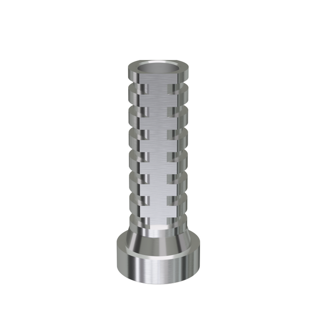
The structural integrity of an implant-supported prosthetic relies on the connections between the components. The Titanium Base (ti-base) abutment plays a major role in modern implant dentistry. It serves as the precision interface between the surgical implant fixture and the final prosthetic framework. The ti-base ensures a passive fit, which is essential for preventing stress accumulation in the restorative material. It prevents micro-leakage of bacteria into the implant connection and absorbs occlusal shock. Utilizing a precision-milled titanium base provides vastly superior mechanical stability compared to direct-to-implant restorative connections, significantly reducing the risk of screw loosening or framework fracture.
Pink Esthetics (Soft Tissue Replication)
Severe tooth loss is always accompanied by the resorption of the alveolar bone and the collapse of gingival tissue architecture. Advanced full-arch restorations utilize pink acrylic or pink zirconia to reconstruct this lost volume. This "pink esthetics" approach restores correct facial profile support, plumping the lips and cheeks. It also prevents the formation of "dark triangles" near the lip line, ensuring a natural transition between the prosthetic and the patient's remaining tissues.
Surgical Realities and Prerequisites
Full-arch implant reconstruction requires adequate bone volume to anchor the titanium fixtures. If bone resorption is severe, preparatory procedures such as bone grafting or sinus lifts may be necessary before implant placement. Patients must also understand the reality of the osseointegration healing phase. While provisional teeth are often attached on the day of surgery, the internal integration of the implants with the jawbone takes three to six months. During this period, strict adherence to a soft diet is mandatory to prevent micro-movement and implant failure.
Evaluation Dimensions: Matching the Prosthetic to the Patient
Selecting the appropriate prosthetic requires a comprehensive evaluation of the patient's specific anatomical and functional circumstances. A one-size-fits-all approach inevitably leads to suboptimal outcomes. We must look at the mechanical forces at play in the patient's mouth.
Biomechanical and Functional Demands
Parafunctional habits, specifically bruxism (teeth grinding) and clenching, dictate material selection. A patient with severe bruxism generates occlusal forces that will easily fracture layered porcelain or acrylic. For these individuals, steering toward monolithic zirconia over layered ceramics is a clinical necessity. The prosthetic design must also incorporate specific occlusal schemes to minimize lateral interferences during jaw movement, protecting both the restoration and the underlying implants or natural teeth. We map the bite forces using articulating paper and digital occlusal analysis to ensure even force distribution.
Scalability and Future-Proofing
Treatment planning should account for progressive tooth loss. A strategic approach involves future-proofing the restorations. For example, a single implant placed today can later serve as an abutment for a bridge if the adjacent tooth fails. A failing dentition can be transitioned to a full-arch prosthetic in stages to manage surgical impact. Utilizing universally compatible implant systems and modular prosthetic designs allows clinicians to adapt the restoration as the patient's oral health evolves over decades.
Implementation Risks and Mitigation Strategies
All prosthodontic interventions carry inherent risks. Understanding these complications and the strategies used to mitigate them is essential for patient informed consent and long-term success. We do not ignore the possibility of failure; we engineer protocols to prevent it.
Surgical Complications
Implant placement involves surgical risks. Peri-implantitis, an inflammatory condition affecting the tissues surrounding an implant, can lead to rapid bone loss and implant failure if left untreated. Failure of osseointegration occurs when the bone does not fuse to the titanium surface, often due to micro-movement during healing or underlying systemic conditions. Nerve proximity issues, particularly concerning the inferior alveolar nerve in the lower jaw, can result in temporary or permanent altered sensation if the implant is placed too deeply.
Prosthetic Failures
The restorations themselves are subject to mechanical failure. Porcelain chipping is common in layered restorations subjected to heavy bite forces. Screw loosening in fixed implant prosthetics can occur if the framework lacks a passive fit or if the occlusal forces are unbalanced. Cement wash-out in traditional crowns and bridges allows bacteria to infiltrate the margins, leading to rapid, undetected decay of the underlying tooth structure.
Risk Mitigation
Modern clinical protocols heavily emphasize risk mitigation. The use of 3D Cone Beam Computed Tomography (CBCT) imaging is non-negotiable for surgical planning, allowing the clinician to map bone volume and nerve pathways in three dimensions. Surgical guides, 3D-printed based on the CBCT data, ensure precise implant trajectory and depth. Strict adherence to post-operative soft-diet protocols during the provisional phase prevents premature loading and micro-movement, securing the osseointegration process.
Conclusion
Selecting the appropriate prosthetic requires a comprehensive evaluation of the patient's specific anatomical and functional circumstances. A one-size-fits-all approach inevitably leads to suboptimal outcomes. Your choice between single-tooth crowns, partial arch bridges, or comprehensive full-arch implant restorations must be dictated by remaining tooth structure, periodontal health, bone volume, and the patient's long-term functional demands.
As a premier global innovator in premium medical devices and high-precision restorative engineering, KETAI develops and manufactures advanced CAD/CAM restorative components, clinical dental materials, and specialized prosthodontic solutions tailored to meet the strict biocompatibility and durability standards of modern implantology. By combining advanced metallurgy with micron-level machining precision, KETAI equips dental laboratories and clinical institutions with premium titanium bases, multi-unit abutments, and modular restorative systems that eliminate structural stress concentration and optimize long-term clinical outcomes.
FAQ
Q: How long do dental prosthetics typically last?
A: Traditional crowns and bridges have an average lifespan of 10 to 15 years, often requiring replacement due to secondary decay or marginal failure. Implant-supported prosthetics possess a 20+ year potential, frequently lasting a lifetime, as titanium implants are impervious to decay. Longevity for all prosthetics is heavily contingent on strict daily hygiene and regular professional maintenance.
Q: What is the difference between a crown and a bridge?
A: A crown caps and encases a single existing, damaged tooth to restore its structural integrity and shape. A bridge is used to replace one or more completely missing teeth. It spans the edentulous gap by anchoring a false tooth (pontic) to the adjacent natural teeth or to dental implants.
Q: When does a tooth need a dental crown instead of a dental filling?
A: A filling patches a small to moderate cavity where the surrounding enamel remains strong. A crown is required when structural loss exceeds 50% of the tooth volume. At this stage, the remaining tooth structure cannot support normal biting forces and risks catastrophic vertical fracture without the full-coverage protection of a crown.
Q: Are implant-supported prosthetics better than traditional dentures?
A: Yes, functionally and biologically. Implant-supported prosthetics preserve the jawbone by providing internal stimulation, preventing facial collapse. They restore over 90% of natural bite force, allowing patients to eat a normal diet, and completely eliminate the slipping, clicking, and mucosal irritation associated with traditional removable dentures.
Q: How does a titanium base improve full arch restorations?
A: A titanium base acts as the precision-machined interface between the surgical implant and the final prosthetic framework. It provides a highly accurate, durable connection that ensures a passive fit. This reduces stress on the restorative materials, prevents micro-leakage, and significantly lowers the incidence of mechanical failures like screw loosening.
Q: What is the difference between dental veneers and dental crowns?
A: Veneers are thin, aesthetic shells of porcelain bonded only to the front surface of anterior teeth, requiring minimal enamel preparation. They alter shade and shape but offer little strength. Crowns encase the entire tooth circumferentially, requiring significant preparation, to restore heavy mechanical strength and function.
Q: What is the recovery time for full arch dental implants?
A: Initial soft tissue healing of the gums typically takes 1 to 2 weeks, during which patients manage minor swelling and discomfort. However, the internal osseointegration process—where the bone fuses to the titanium implants—takes 3 to 6 months. A strict soft diet is required during this phase to ensure success.